Ultrasound test
The advancements in medical technology have truly enhanced our lives by enabling early diagnosis and treatment of diseases and medical conditions. One innovation in this field is the Ultrasound scan, which has significantly improved how we manage a variety of health issues.
Ultrasound test In Australia, Get Tested Today
What:
Real-time scan using sound waves to image internal organs and tissues
Tests for:
Abdomen and thyroid abnormalities
Referral:
Required
Member cost:
$200 - $1000
What is an Ultrasound scan?
An ultrasound scan is an imaging method that creates a real-time picture of what’s happening inside the body using sound waves. Ultrasound is typically painless and non-invasive, making it a comfortable choice for many individuals. Unlike X-rays, ultrasound is completely free of radiation, ensuring your health remains a top priority.
What are the types of Ultrasound scan?
The two categories of Ultrasound include diagnostic and therapeutic.
Diagnostic ultrasound
Diagnostic ultrasound is a non-invasive technique that helps generate images of what’s happening inside the body. Using special devices called transducers, it sends out sound waves at frequencies in the megahertz range, much higher than what you can hear (above 20 kHz). Most diagnostic ultrasound probes are simply placed on the skin or gently inserted into the body through areas like the gastrointestinal tract or blood vessels. It can be further subdivided into:
- Anatomical ultrasound: It produces images of internal organs such as the liver, pancreas, stomach, kidneys, prostate, uterus, thyroid or other structures such as muscle tendons, skin layers
- Functional ultrasound: It integrates tissue movement, velocity, softness, and other traits, creating "information maps.” These maps help doctors visualise changes and differences in organ function. For instance, Doppler ultrasound measures the speed and intensity of blood flow in the blood vessels
Therapeutic ultrasound
Therapeutic ultrasound uses sound waves beyond human hearing, interacting with body tissues to modify or alter them. Possible modifications include moving tissue, heating, dissolving blood clots, delivering drugs, and destroying diseased tissues such as tumours.
What are the uses of abdominal and thyroid ultrasound?
Abdominal ultrasound
Your doctor will recommend doing an abdominal ultrasound to evaluate the internal organs in your abdomen. It is used to:
- Show a picture of internal organs such as liver, gallbladder, bile ducts, pancreas, spleen, kidneys, abdominal aorta and check their size, shape and position
- Determine the cause of abdominal pain, such as gallstones, kidney stones, trauma
- Identify cysts, tumours, fluid collections, and other abnormalities within the abdominal cavity
- Screen and monitor aneurysms in the aorta
- Assist in positioning needles for biopsies or draining fluid collections
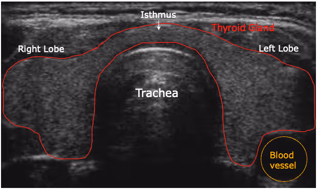
Thyroid ultrasound
Your doctor will recommend doing a thyroid ultrasound to assess the thyroid gland, which helps in knowing if it is functioning correctly. It is used to:
- Identify the size and shape of the thyroid gland
- Monitor the size of the thyroid gland during treatment
- Identify thyroid nodules and assess their size, shape, quantity, and whether they are benign or malignant
- Assess enlarged parathyroid glands, commonly linked to various conditions
- Assist in positioning a needle during a biopsy to collect a sample from a nodule for additional examination
How does Ultrasound work?
Ultrasound waves are created by a transducer that not only emits these waves but also detects the echoes that bounce back. Typically, ultrasound transducers are composed of special ceramic crystal materials known as piezoelectrics. These materials can generate sound waves when an electric field is applied to them, and they can also work the other way around, producing an electric field when sound waves reach them.
In an ultrasound scanner, the transducer works by sending a gentle beam of sound waves into the body. As these sound waves travel, they bounce back to the transducer when they encounter different tissue boundaries, like the ones between fluid and soft tissue or between tissue and bone. When the echoes reach the transducer, they create electrical signals that are sent to the ultrasound scanner. By measuring the speed of sound and timing how long it takes for each echo to return, the scanner can calculate the distance from the transducer to the tissue boundary. These distances are then transformed into two-dimensional images of our tissues and organs.

How do you prepare for an ultrasound scan?
To prepare for an ultrasound scan:
- Bring your referral letter and any ultrasound scan or X-ray results you have received over the past 2 years, if they were performed at another location
- Wear comfortable, loose-fitting clothing
- Leave your jewellery and valuables at home
- The abdominal ultrasound test has specific instructions for eating and drinking:
- Before an ultrasound of the liver, gallbladder, spleen, and pancreas, you may need to have a fat-free meal the night before and refrain from eating for 8-12 hours before the test
- Before the kidney ultrasound, you might need to drink four to six glasses of liquid approximately one hour prior to the test to ensure your bladder is full. Additionally, you may be instructed to refrain from eating for 8-12 hours before the test to minimise gas accumulation in the intestine
- For ultrasound of the aorta, you may need to avoid eating for 8 to 12 hours before the test
- No special preparation in terms of eating or drinking is required for a thyroid ultrasound
What occurs during an ultrasound?
An ultrasound test usually takes 30 minutes to an hour. Preparation for an ultrasound varies depending on the body part being scanned. Your provider may ask you to remove certain pieces of clothing or change into a hospital gown.
Ultrasounds that involve applying the transducer (probe) over your skin (not in your body), follow these general steps:
- You can relax on your side or back on a cosy table, making yourself comfortable
- The ultrasound technician will gently apply a small amount of water-soluble gel on your skin in the area that needs to be examined. This gel is safe for your skin and won’t stain your clothes
- The technician will gently move a handheld transducer or probe over the warm gel to capture clear images of what's happening inside your body
- The technician may ask you to remain still or hold your breath for a few moments to help create clearer images
- After the technician has collected sufficient images, they will remove any leftover gel from your skin, and then you will be finished

Post-Test Care or What to Expect After the Test
Resumption of Normal Activities
You can jump straight back into your day once you leave the clinic. Whether it’s returning to work, hitting the gym, or making dinner, there’s no recovery time needed. Just carry on as usual.
Possible Mild Discomfort
You might feel a little tenderness or warmth where the probe pressed against your skin—especially if gentle pressure was needed for clearer images. This mild sensation usually eases within minutes to a couple of hours. If it lingers, a cool pack or simple over-the-counter pain relief should sort it.
Results Timeline
Your scan images go to a radiologist for review, and you’ll typically get the report within 24–48 hours. Your referring doctor then goes over the findings with you. If you’ve set up an online health portal, you may even spot a notification or preliminary notes before your follow-up chat.
Follow-Up Appointments
Depending on what the radiologist finds, your doctor may book a follow-up to discuss next steps—this could involve more imaging, a specialist referral, or routine monitoring. If anything urgent turns up, you’ll hear back straight away. Otherwise, expect to hear about any recommended actions within a week.
When can you expect to receive the results of your ultrasound?
The time it takes to receive your results can vary a bit depending on the type of ultrasound you have. A radiologist will carefully analyse the images and send the report to the doctor who requested the exam. Your doctor will then share the results with you, or you might find them available in your electronic medical record if you have an account set up even before your doctor has a chance to review everything with you.
Sometimes, based on your symptoms, your doctor may recommend an urgent ultrasound of your abdomen or thyroid. In such cases, your doctor will notify you about the results and further management on an urgent basis.
Interpretation of Ultrasound Results
Abdominal ultrasound
A radiologist may indicate that the findings in different abdominal organs are normal or abnormal. Common abnormal findings may include cysts or calcifications in the pancreas, fatty liver, gallstones, kidney stones, or hydronephrosis.
For an abnormal finding, the radiologist may recommend:
- Additional imaging tests that can help better assess the finding or obtain a follow-up imaging test to re-evaluate the finding after some time
- Biopsy
- Correlating the findings with clinical symptoms or laboratory test results
- Comparing the findings to other imaging studies that the radiologist interpreting your test cannot access. This often occurs when your imaging tests are conducted at various facilities or hospitals

Thyroid ultrasound
Similar to abdominal ultrasound, the findings on the thyroid ultrasound are described as normal or abnormal. Abnormal results may be due to cysts, enlargement of the thyroid gland, thyroid nodules, thyroiditis, or inflammation of the thyroid and thyroid cancer.
The abnormal findings include irregular margins, microcalcifications, height greater than width, extension outside the thyroid, and cervical lymph nodes with suspicious features. Many thyroid ultrasound reports will now assign a score to each nodule and describe the characteristics that influenced the score.
Depending on the findings, the radiologist recommends a further course of management. Your doctor follows this in correlation with the presenting symptoms.


What are the risks involved with an ultrasound?
Diagnostic ultrasound is widely viewed as a safe option since it does not produce ionising radiation like X-rays do. However, it's important to note that ultrasound can create some biological effects in the body under certain conditions. Because of this, the FDA has set guidelines to ensure that diagnostic ultrasound devices work within safe limits. Additionally, the FDA and many professional organisations advise against using ultrasound casually, such as for keepsake videos, and suggest that it should only be performed when there's a genuine medical need.
What are the limitations of ultrasound?
Ultrasound is generally not the preferred option for imaging bones or air-filled tissues, such as the lungs. However, under certain conditions, it can effectively capture images of bones, particularly in foetuses or small babies, and also the lungs and their lining when they contain fluid, either fully or partially.
Ultrasound might be a bit less effective for those who are living with overweight or obesity, as the sound waves have to travel through more fat to get to the target area.
What is the ultrasound test price?
Ultrasound services marked with the symbol ‘R’ are only eligible for a Medicare ultrasound coverage benefit when they are performed under professional supervision.
Supervision may come from either:
- A specialist in the practice of their speciality
- A consultant physician in the practice of their speciality
An ultrasound service can be supervised by a practitioner who isn’t a specialist or consultant physician, as long as they meet the requirements outlined in Note IN.0.13 on the MBS Online website.
Practitioners have the opportunity to claim Medicare ultrasound coverage benefits for ultrasound services when they are provided in an emergency, or if the location is more than 30 kilometres away via the most direct road route from another practice. This ensures that patients receive the care they need, irrespective of their location.
Sources
1. National Institute of Biomedical Imaging and Biomedical Engineering - Ultrasound.
https://www.nibib.nih.gov/science-education/science-topics/ultrasound
2. Cai L, Pfob A. Artificial intelligence in abdominal and pelvic ultrasound imaging: current applications. Abdom Radiol (NY). 2025;50(4):1775-1789. doi:10.1007/s00261-024-04640-x
3. Xie C, Cox P, Taylor N, LaPorte S. Ultrasonography of thyroid nodules: a pictorial review. Insights Imaging. 2016;7(1):77-86. doi:10.1007/s13244-015-0446-5
4. Radiologyinfo.org - Abdominal Ultrasound.
https://www.radiologyinfo.org/en/info/abdominus
5. Mount Sinai - Thyroid ultrasound.
https://www.mountsinai.org/health-library/tests/thyroid-ultrasound
6. Radiologyinfo.org - Thyroid Ultrasound.
https://www.radiologyinfo.org/en/info/us-thyroid
7. Services Australia - Ultrasound services.
https://www.servicesaustralia.gov.au/mbs-billing-for-ultrasound-services?context=20
Conclusion
Ultrasound is a safe, effective, and radiation-free way to understand what’s going on inside your body—whether it’s checking your thyroid, assessing abdominal pain, or guiding treatment decisions. It's non-invasive, widely available across Australia, and often covered by Medicare when performed under the right supervision.
Additional FAQs


imaging
biomarkers
sterols
Book a Free Discovery Call
Join 20,000+ Australians improving their health with proactive, personalised healthcare.